What’s involved in a 3D ultrasound?
Having a 3D ultrasound is not such a different process to having a 2D ultrasound, at least for the mother. However, although the procedures are much the same, they use very different levels of technology to provide very different images.
A 3D ultrasound takes thousand of pictures or photos of the baby at the once time. These are then translated by computer into 3 dimensional images which are almost as clear as a real life photograph. These “still” pictures of your baby mean that you can see your baby in three dimensions, rather than two.
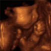
There is a depth and shape to a 3D image, giving a clarity which is not as evident in a 2D ultrasound. This is because a 2D ultrasound sees through the baby to its internal organs and tissues. Whereas, with a 3D and 4D ultrasound the baby’s skin can be seen so there is more of a realistic shape and form to the images. This is particularly clear when looking at the baby’s face and delicate features.
With 3D ultrasounds, both the transducer used to transmit the sound waves and the computer software are more advanced and complex. This is why a 3D ultrasound is more expensive and generally not included in standard obstetric practice.
How are 2D and 3D ultrasounds different?
A 2D ultrasound takes image “slices” which can only be viewed by looking at one image at a time. Sound waves are sent to the baby and reflected straight back which means there is a rather flat, surface appearance to the baby. Whereas a 3D ultrasound involves taking thousands of “slices” in a rapidly occurring series called a “volume of echoes”. These send sound waves back at different angles, allowing for the characteristic 3D depth.
Once these pictures are stored and shaded by the computer, they can be seen on the screen as clearer, 3 dimensional images. The width, height and depth of the baby and its internal organs can be seen very clearly. With 3D ultrasound the baby has a more realistic shape and form, with distinctly baby type features and look exactly as they would if they were already born, only smaller of course.
Parents need to rely less on their imagination with a 3D ultrasound. It’s as if all the 2D images have been filled in and puffed out so the image is quite clearly a baby, rather than a grainy image on the screen.
What’s the benefit of having a 3D ultrasound?
Although it’s lovely to see the baby with some a clear image, there are no real health benefits to having a 3D ultrasound when compared to a 2D one. Since the late 1990s, 3D ultrasound has been available and thousands of parents have elected to have this done. Specialist obstetric scanning clinics provide 3D technology and parents need to pay to have this procedure.
Free public hospital and Medicare rebated scans are generally limited to 2D ultrasounds because of the additional cost of the 3D technology.
Taking a 3D ultrasound requires a high level of clinical skill and expertise. Unlike the 2D ultrasound, a 3D ultrasound requires the transducer to be held still and steady whilst the sound echoes bounce back and are then interpreted by the computer software. If you have had a 2D ultrasound previously you will find the imagery very different.
When is the best time for me to have a 3D ultrasound?
Generally, the recommendation is between 26-30 weeks gestation, unless otherwise suggested by your maternity care provider. By this time, there is sufficient fat under the skin to see the baby’s facial appearance, rather than the supportive bony structure.
Some clinics claim that past 30 weeks of gestation, the baby’s head is more likely to engage in the mother’s pelvis so visualising the face can be more difficult. But this really depends on the individual clinic and their own practice guidelines.
How long will it take to have my 3D ultrasound?
A 3D ultrasound tends not to take as much time to complete as a 2D. This is because the images are so clear and they can be stored within the computer’s hard drive for later use if necessary. Sonographers aim to finish doing the 3D ultrasound within 30 minutes of starting the procedure. This is to limit any potential side effects on the baby from being exposed to prolonged sound waves.
But I can’t see a thing!
How much you see of your baby during the 3D ultrasound will really depend on the way they are laying. If your baby is facing outwards and there is enough amniotic fluid surrounding their little face, then you will be in luck. Even so, you will be able to see their back, shoulders, bottom and little limbs.
If, at the beginning of the scan your baby is facing away from the transducer, keep your fingers crossed that may move or rotate into a clearer position before the scan is finished. If your baby is curled up tightly, facing your back or has their hands covering their face, then you won’t be able to see much of their features.
If you are carrying a lot of weight around your middle, this could influence the clarity of the image you see. The sonographer may suggest you get up and go for a little walk or have a cold drink, talk to your baby or gently massage your abdomen. These strategies may help to encourage your baby to move into another position.
Options available at some 3D ultrasound clinics
- You may be given the option of taking home photos or a video of your 3D ultrasound. Check with the receptionist when you are making your appointment if you need to bring a memory stick with you.
- You may be able to invite family members to view the ultrasound with you. Some clinics have movie theatre style seating arrangements so the experience can be shared.
- Your 3D movie can be set to the music of your choice.
- You may be able to take home some photos of your scan.
Benefits of having a 3D ultrasound compared to a 2D
There are no definite medical benefits; however, if a defect has been identified a 3D ultrasound may provide better visualization than a 2D ultrasound . This may apply to issues such as a heart defect, cleft lip or a neural tube defect – for example spina bifida. Because of this early detection and awareness, planning for birth, postnatal care and management can begin very early. It can also help parents to know what they may be faced with when their baby is born, rather than needing to rely on their imaginations and the explanations of healthcare professionals.
Some parents find that having 3D ultrasound helps them to bond with their baby before birth. Having a clear view of their baby’s face and features, looking for family likenesses, knowing the baby’s gender and being able to see their baby makes a big difference.
At the time of the 3D ultrasound, some parents choose to name their baby and see this as a unique opportunity to build early emotional attachment. Yet for others, they are happy to wait until when their baby is born to meet for the first time.
What’s the difference between a 3D and 4D ultrasound?
4D or 4 dimensional scans build on the technology of 3D scans. The extra dimension is time, so that effectively 4D ultrasounds are moving images of your baby in real time. As your baby kicks, moves, frowns, grimaces, sucks their thumb, opens their eyes and moves their lips, you will be able to see these movements on the scan as they are actually happening.
The technology associated with 4D ultrasounds makes them more expensive and they are not generally offered as part of a routine obstetric service. If parents choose to have a 4D ultrasound done, they will need to pay for this procedure themselves. If it is done in a medically aligned sonography practice, Medicare may rebate some of the fee. Alternately, some private health insurance companies could provide a rebate. Check with your individual provider.
3D Ultrasounds are also known as elective prenatal ultrasounds. They are not designed to replace the standard 2D procedure which is commonplace obstetric procedure. Instead they offer a more enhanced “bonding” experience with the unborn baby and are instigated by parents choosing to have them done, rather than a medical need.
Some parents are very keen to see for themselves how their baby is growing and developing, and for this reason another name for the 4D ultrasound is a “lifestyle” scan.
Important information
Both 3D and 4D ultrasounds are not designed to replace a standard obstetric ultrasound. Many companies will not do a 3D or 4D ultrasound unless a 2D medical diagnostic ultrasound has been done first. This is to screen out any abnormalities for both the mother and her baby.
Mothers also need to be engaged with a pregnancy healthcare provider first before having a 3D or 4D ultrasound.
How much will a 4D ultrasound cost?
Since technology has expanded and 3D/4D scans have become readily available, there is a lot of competition in the market. Different clinics claim to provide superior and more enhanced images and their advertisements can be very enticing.
At the non-diagnostic clinics, packages range in cost from around $100-$200 depending on what level of service you want. You can get a DVD with or without music of your choice, image print-offs and a CD of the images.
Some practices have special discounted prices, when couples make appointments for more than one 4D ultrasound session. Ideally, these are some months apart. For couples who are keen to gauge the individual growth and development of their little baby, these 4D ultrasounds can be fascinating.
When should I have my 4D ultrasound?
There is some discrepancy between clinics around when a 3D and/or 4D ultrasound should be done. Many claim the optimum time is somewhere between 27-32 weeks of gestation and others state between 26- 30 weeks. On either side of these time frames a three week “window” will still provide a good viewing alternative.
From 19-24 weeks of gestation, gender determination scans can be done. At this early stage though, the baby does not have enough fat to “fill out” its facial features, so the quality of the scan and images you’ll see won’t be as clear as they will be later. Waiting for just a little longer and being patient will reap a lot of benefits!